
Figure 7.
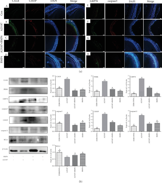
BMP4 relieved ER stress through CALR in chicken SWFs. The sections of the SWFs were immunofluorescent labeled ((a) A–D), and the ER stress marker CALR (green) and the ER stress apoptosis transcript CHOP (red) were mainly distributed in the GL, and a small amount was distributed in the TL. Histological sections of SWFs were given immunofluorescent labels ((a) E–H) with ER stress marker GRP78 (green) and apoptosis marker caspase3 (red), and they were coexpressed in the GL. Scale bar: 50 μm. Western blot and grey analysis (b) of CALR, PERK, GRP78, caspase3, CHOP, caspase12, and BCL2. Values were the mean ± SEM of three experiments. Different lowercase letters indicated significant difference (p < 0.05).