
Figure 3:
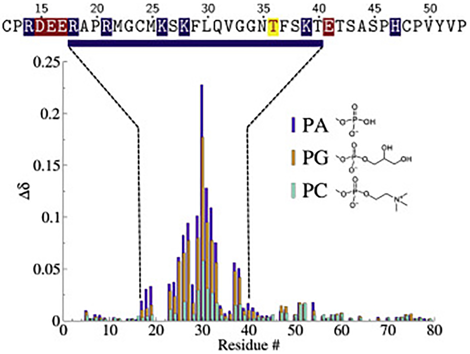
Chemical shift perturbations (CSPs, Δδ = [(Δδ1H)2 + (Δδ/515N2)]) of HckSH4–U with PA (purple), PG (orange), and pure PC (teal) bicelles. The sequence region experiencing larger CSPs is displayed above the data and highlights acidic (red) and basic (blue) residues near the lipid binding span. The enriched basic character spanning R18 to T40 suggests that the binding to negatively charged lipids is driven by electrostatics. The location of the phosphorylation site Thr36 is marked in yellow.