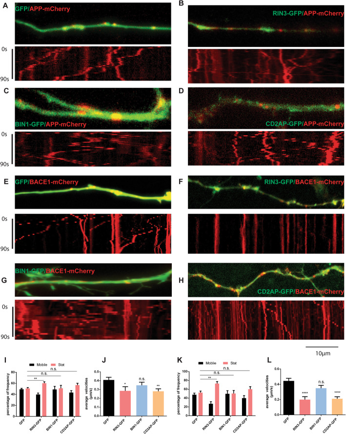
Fig. 6.
RIN3 and CD2AP inhibited transport of APP and BACE1 in primary cortical neurons. E18 mouse cortical neurons were cultured in microfluidic chambers and were co-transfected with the indicated expression vectors. Live imaging was performed as described in the Methods section. Representative images of axons and corresponding kymographs are shown in a-h. The percentiles of mobile versus stationary vesicles (stat) (i, k) and average velocities (g, l) for APP-mCherry and BACE1-mCherry in axons were quantitated. Data represent mean ± SEM of at least 3 independent experiments. All p-values were calculated using 1-way ANOVA. p < 0.05 (*), p < 0.01(**), p < 0.0001(****), p > 0.05 (n.s.), standard t-test

