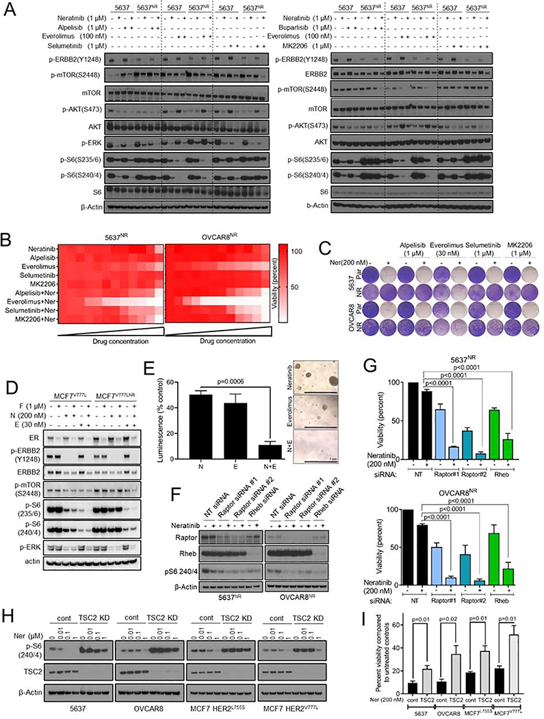
Fig. 3. Genetic and therapeutic suppression of TORC1 overcomes resistance to neratinib.
(A) Immunoblot analysis of parental and neratinib-resistant 5637 cells treated with the indicated drugs combinations for 24 hr; alpelisib (PI3Ka inhibitor), everolimus (TORC1 inhibitor), selumetinib (MEK1/2 inhibitor), buparlisib (pan-PI3K inhibitor), MK2206 (AKT inhibitor). (B) Heatmaps representing 12-point dose response assays of 5637NR and OVCAR8NR cells treated with the indicated single agents or combinations. For single agents, cells were treated with increasing concentrations (3-fold) of the drug, up to 1 μM. For combination assays, all cells were treated with 1 μM neratinib and increasing concentrations of the second drug. (C) Representative images of cells seeded in 12-well plates, treated with the indicated drug combination; drugs and media were replenished every 72 hr. When control cell monolayers reached ~90% confluency, they were stained with crystal violet and imaged. (D) Immunoblot analysis of parental and neratinib-resistant MCF7V777L cells treated with the indicated drugs for 24 hr. (E) Growth of organoids established from neratinib-resistant ST1616B (HER2D769Y) breast cancer PDX. Organoids were treated with vehicle, neratinib (100 nM), everolimus (30 nM) or the combination. Viability was assessed 6 days later by measuring ATP levels and normalized to vehicle treated control. Values are mean ± SEM from three independent experiments, Student’s t-test. Representative images of organoids from each treatment group are shown on the right, scale bar = 1 mm. (F) Immunoblot analysis of neratinib treated 5637NR and OVCAR8nr cells transfected with the indicated siRNAs. (G) Viability of neratinib-treated cells transfected with the indicated siRNAs. When control cells reached ~90% confluency, cells were trypsinized and counted using a Coulter counter. Values are mean ± SEM from three independent experiments, one-way ANOVA. (H) Immunoblot analysis of lysates from 5637, OVCAR8, MCF7L755S and MCF7V777L cells stably transfected with TSC2 shRNA or empty-vector control. Cells were treated with increasing concentrations of neratinib; 24 hr later, cell lysates were prepared and tested by immunoblot with the indicated antibodies. MCF7L755S and MCF7V777L cells were tested under estrogen-free conditions. (I) Viability of 5637, OVCAR8, MCF7L755S and MCF7V777L cells transfected with TSC2 shRNA or empty vector and treated with neratinib. Drug and media were replenished every 72 hr. MCF7 and MCF7V777L cells were treated with neratinib under estrogen-free conditions. When control cells reached ~90% confluency, monolayers were trypsinized and cell number measured using a Coulter counter. Each bar represents mean cell viability ± SEM from three independent experiments, Student’s t-test. See also Figure S3.

