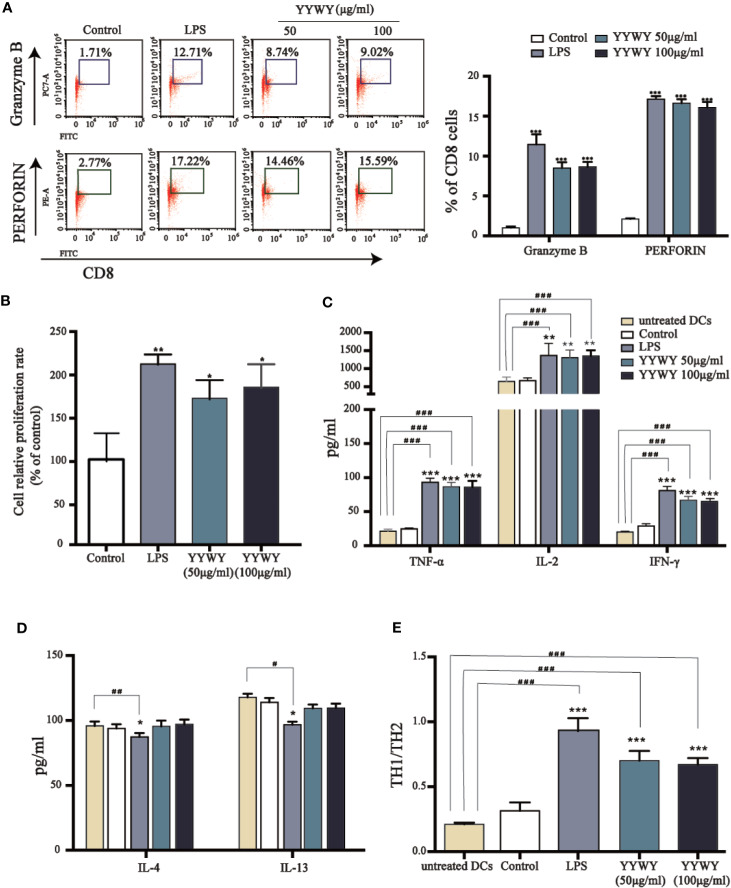
Figure 5.
YYWY enhanced DCs-induced proliferation and programming of T cells. BMDCs were treated with YYWY (50 or 100 μg/ml) for 48 h, followed by collecting the DCs for subsequent co-culture with allogeneic CD3+ T cells for another 48 h. (A) The detection of PERFORIN and GRANZYME through flow cytometry was used to assess the differentiation of CD3+ T cells. Representative FACS analysis scatter grams are presented on the left. The quantification of PERFORIN and GRANZYME-positive cells which were normalized to total CD3+ T cells is presented on the right. (B) The proliferation of CD4+ T cells was estimated by CCK-8 assay. (C–E) The cytokine levels of Th1 type cytokines (IL-2, TNF-α, IFN-γ), Th2 type cytokines (IL-4, IL-13), and Th1/Th2 (IFN-γ/IL-4) radio were detected by Elisa assay. The values represent the mean ± SEM of three independent experiments. *p < 0.05, **p < 0.01, ***p < 0.001 compared to the control group, #p < 0.05, ##p < 0.01, ###p < 0.001 compared to the untreated DC.

