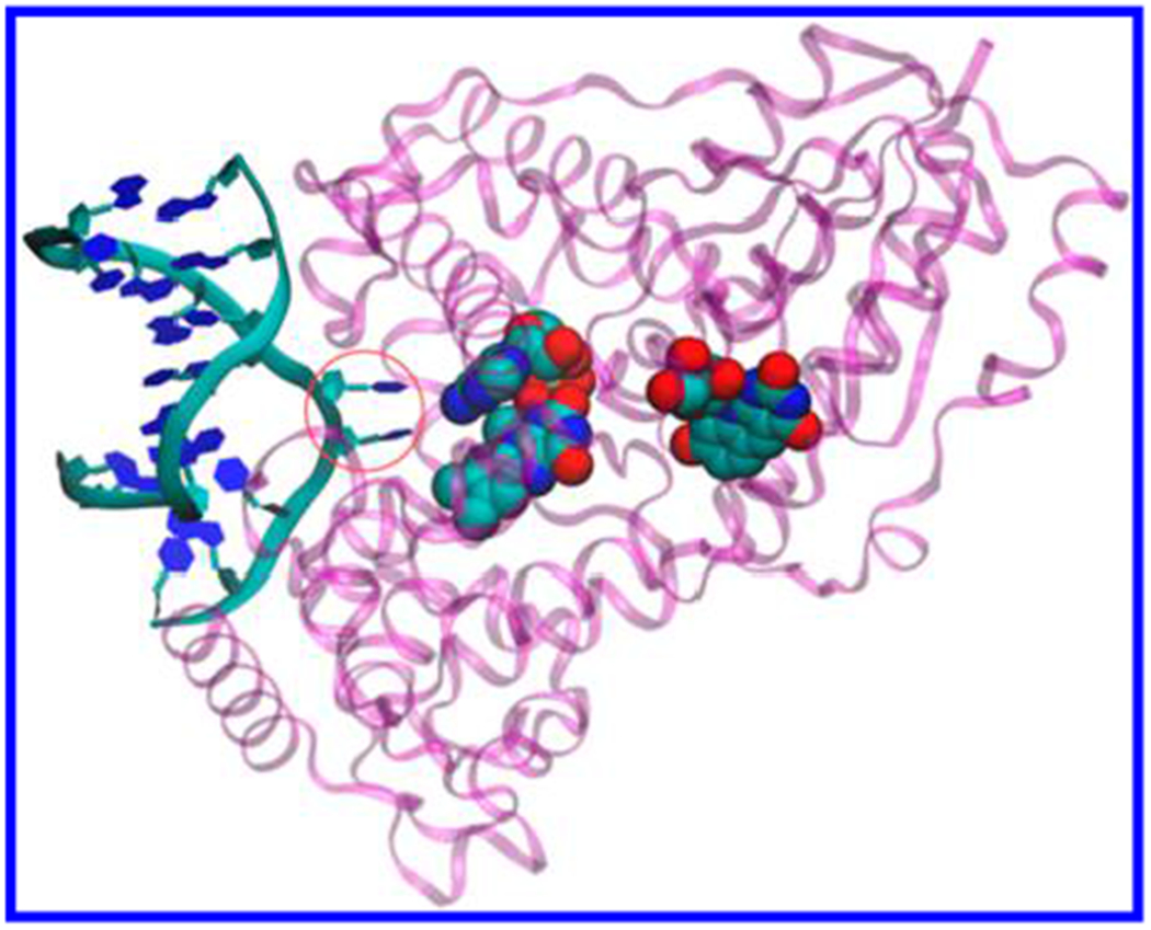
Figure 1.

DNA photolyase from A. nidulans (purple) complexed with repaired DNA analog (left) (crystal structure from the PDB file 1TEZ20). ET from the excited FADH− cofactor (CPK representation, center) splits the two thymines in CPD (circled blue hexagons). The 5-deazaflavin antenna cofactor is also shown (CPK representation, right).
