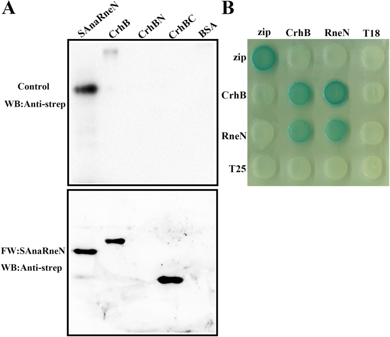
FIG 3.
Identification of the CrhB interaction domain in AnaRne. (A) Far-Western blotting assays designed to determine the regions of AnaRne that interacted with CrhB. One of the PVDF membranes was incubated with SAnaRneN (bottom), and the other was incubated with skim milk as a control (top). SAnaRneN served as a positive control, while BSA served as a negative control. The other proteins were purified with the C-terminal His tag. The final results were also detected with the anti-strep antibody. The amount of proteins loaded was 1 μg, except for the positive controls (20 ng). (B) Bacterial two-hybrid analysis of the interaction between AnaRneN and CrhB. “RneN” represents AnaRneN. The horizontal rows represent the E. coli BTH101 cells carrying the recombinant “T18” protein fusion plasmids, whereas the vertical columns represent the cells carrying the recombinant “T25” protein fusion plasmids. These results were based on three independent experiments.

