Fig. 1.
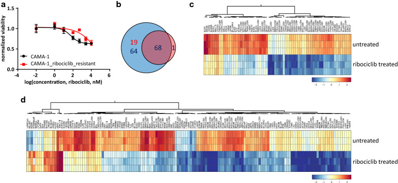
Transcriptional response to ribociclib in sensitive and ribociclib-resistant cells. a Dose–response curves of CAMA-1 and CAMA-1_ribociclib_resistant cells under different concentrations of ribociclib treatment. Cells were treated with increasing concentration of ribociclib for 96 h, after which viability was measured using CellTiterGlo Chemiluminescent kit. The measured luminescence was normalized to the average of the lowest applied concentration (0.01 nM). Data points show the average of three replicates, error bars show standard deviation if it is larger than the size of the data point. b Venn diagram demonstrating the number of significantly differentially expressed genes in response to 12 h of 1 µM ribociclib treatment in CAMA-1 and CAMA-1_ribociclib_resistant cells. Blue circle incorporates differentially expressed genes in CAMA-1, while the red circle incorporates differentially expressed genes in CAMA-1_ribociclib_resistant cells. Red numbers demonstrate the number of upregulated, while blue numbers demonstrate the number of downregulated genes in response to ribociclib treatment. c, d Heatmaps demonstrating the expression of significantly differentially expressed genes in response to ribociclib treatment in CAMA-1_ribociclib_resistant (c) and CAMA-1 (d) cells

