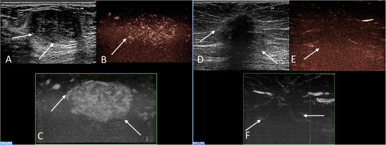
Fig. 1.
Examples of the evaluation of lesions´ vascularity using the MVI technique. Histopathologically verified fibroadenoma in a 37-year-old female patient (a, b, c). There is a well-circumscribed hypoechoic mass (a) with heterogenous internal enhancement (b) and with rich vascularization compared to the surrounding tissue (c). Verified invasive carcinoma of no special type (NST) in a 79-year-old female patient (d, e, f). Greyscale ultrasound shows poorly defined hypoechoic mass (d). A post-processed CEUS image using MVI application displays peripheral penetrating vessels (e) and heterogeneous internal perfusion of the malignant tumor (f). Abbreviations: MVI, MicroVascular Imaging; NST, invasive carcinoma of no special type; CEUS, contrast-enhanced ultrasound

