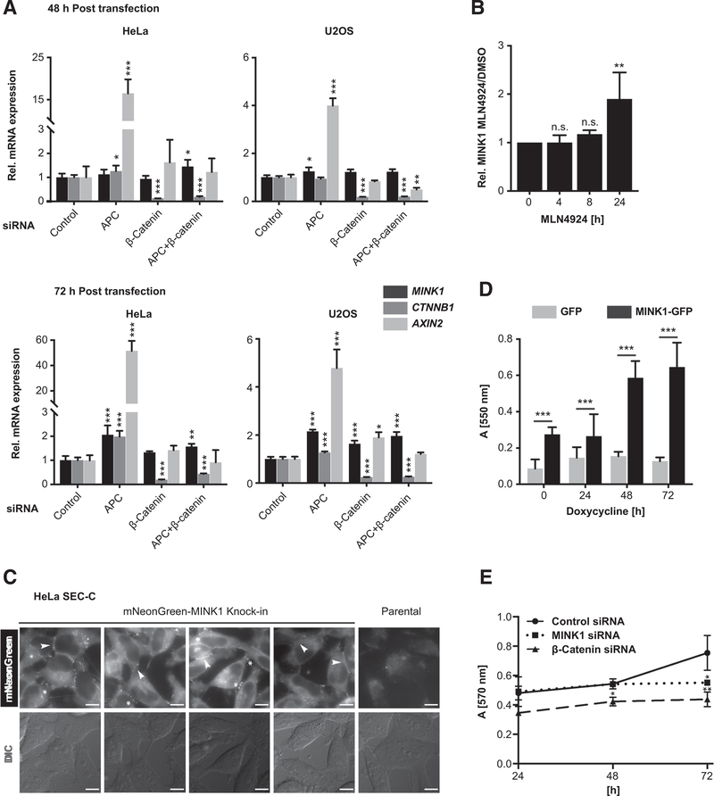
Figure 6.
MINK1 localizes to cell-cell junctions and its overexpression enhances cell adhesion. A, mRNA expression of MINK1, CTNNB1, and AXIN2 measured by qRT-PCR 48 and 72 hours after siRNA transfection. Indicated are mean expression levels relative to ACTB expression with SD from four independent transfections. Significance determined by one-way ANOVA followed by Dunnett multiple comparison test (*, P < 0.05; **, P < 0.01; ***, P < 0.001). Note, the same HeLa AXIN2 mRNA quantification data are also shown in Supplementary Fig. S4D. B, MINK1 protein levels in HeLa cells after treatment with neddylation inhibitor MLN4924 [3 mmol/L] as measured by WB. Shown are relative mean signals normalized to DMSO-treated samples with SD from three independent experiments. Significance determined by one-way ANOVA, **, P < 0.01. C, Live imaging of HeLa SEC-C cells expressing endogenously mNeonGreen-tagged MINK1. Scale bars, 10 μm. D, Adhesion assay with U2OS cells overexpressing MINK1-GFPand GFP, respectively. Adhesion to collagen matrix after 1 hour was quantified by staining of firmly attached cells with Crystal Violet. Indicated is mean absorbance with SD of independent experiments with two different GFP/MINK1-GFP clones and eight technical replicates/condition. Significance determined by two-way ANOVA followed by Sidak multiple comparison test (***, P < 0.0003). E, MTT proliferation assay in Colo320 cells treated with siRNA against β-catenin or MINK1. Shown is the mean absorbance from triplicate measurements. Significance relative to control determined by one-way ANOVA followed by Dunnett multiple comparison test (*, P < 0.05; **, P < 0.01). n.s., not significant.

