
Fig 1. Phosphotyrosine 2D WB signal detected in human lung tumor and normal adjacent tissue samples comigrates with EGFR signal.
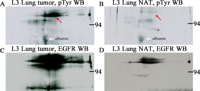
Phosphotyrosine (A and B) and EGFR (C and D) 2D western blots from human lung tumor (squamous cell carcinoma, A and C) and normal adjacent tissue (NAT, B and D) samples purchased from a tissue bank, Bio-IVT. Sample load was 200 μg total protein for each of the four 2D gels. The pTyr-protein signal (red arrows) was strong in the tumor tissue and faint in NAT and co-migrated with the EGFR signal in both. The heavily glycosylated EGFR protein gives a large blurry spot profile due to glycan microheterogeneity. Samples were identically prepared by homogenization in SDS buffer with heating. White arrows indicate nonspecific binding to albumin, an abundant protein that serves as a 2D pI/MW marker.