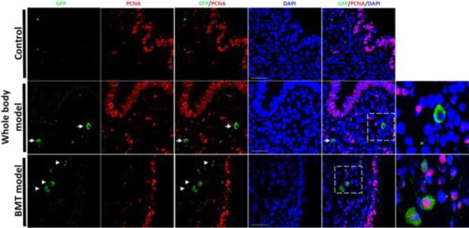
Figure 5.
Proliferation status of fusion-derived cells in mouse endometriosis lesions. PCNA immunostaining of endometriosis lesion sections from control (upper panel), whole body model (middle panel), and BMT model (lower panel). IF photomicrographs demonstrate costaining of GFP-positive fusion-derived cells (green) with PCNA (red). Sections were counterstained with DAPI showing nuclei (blue). White arrows point to GFP-positive cells. Right column: higher magnification of the dashed areas showing GFP-positive cells. Scale bar = 50 μm. n = 12.

