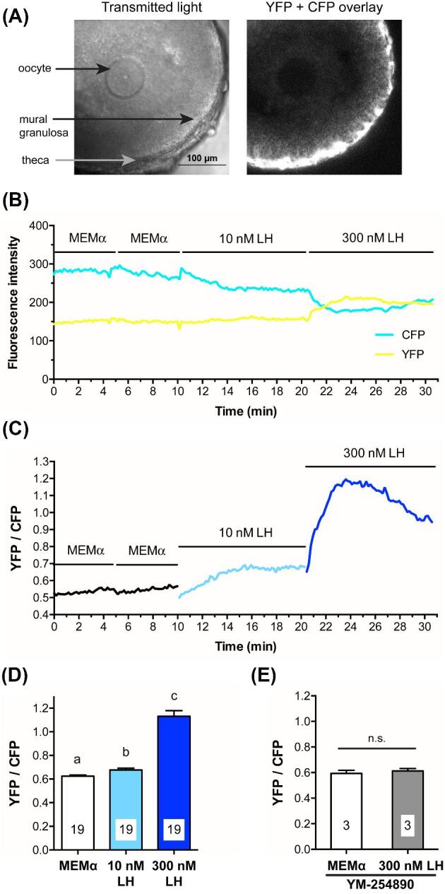
Figure 4.

LH causes a Gq-family G-protein-dependent increase in mural granulosa Ca2+, as detected with Twitch-2B. (A) Transmitted light and CFP + YFP fluorescence images of a large antral follicle after 25-h culture on a Millicell membrane in the presence of 1 nM FSH. Prior to flattening on the membrane, the follicle measured ∼320 μm in diameter. Note that the theca cells express more Twitch-2B than the granulosa cells. (B) YFP and CFP fluorescence for the follicle shown in (A) during sequential perfusions of MEMα, 10 nM LH, and 300 nM LH. (C) YFP/CFP ratio during sequential perfusions of MEMα,10 nM LH, and 300 nM LH, for the follicle shown in B. (D) Peak YFP/CFP ratios before and after perfusion of 10 nM and 300 nM LH. Different letters indicate significant differences (P < 0.001) after repeated measures ANOVA with the Holm-Sidak correction for multiple comparisons. (E) Peak YFP/CFP ratios before and after perfusion of 300 nM LH, following inhibition of Gq-family G-proteins by a 1–2.5 h pretreatment with 10 μM YM-254 890; n.s. indicates P > 0.05 by paired t-test. Values in (D, E) represent mean ± s.e.m.
