
Figure 1.
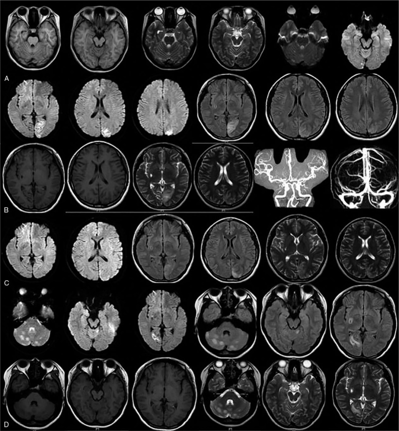
Image data of brain. A: The first hospitalization at 19 years old. Brain MRI showed punctiform long T2 and T1 signals at right cerebellar hemisphere and right occipital lobe, unclear boundary and DWI images with hyperintensity. B: At 21 years old. Brain MRI showed gyrus-shape long T1 and T2 abnormal signals at left-side pillow top and FLAIR and DWI images with hyperintensity. Brain MRV and CTA in head and neck showed no obvious abnormity. C: Reexamination after 10-day treatment. Brain MRI showed that gyrus shape long T1 and T2 signals at left occipital parietal lobe was improved. D: Recurrence at 22 years old. Brain MRI showed schistose and patchy signals at cerebellar hemispheres, vermis cerebelli, and right occipital area. MRI = magnetic resonance imaging.