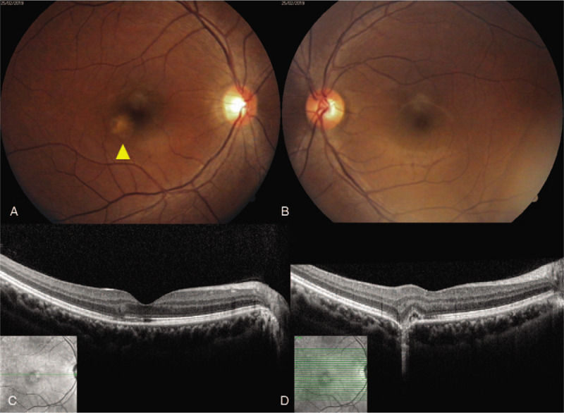
Figure 1.

Fundus photography and optical coherence tomography (OCT) examination at the patient's first visit. (A) The right eye exhibited a yellowish chorioretinal lesion, noted in the parafoveal area. (B) No abnormalities are evident in the left eye. (C) OCT of the right eye revealed focal photoreceptor destruction at the fovea. (D) OCT of the right eye revealed subretinal fluid, destruction of photoreceptors, and retinal pigment epithelium. OCT = optical coherence tomography.
