
Figure 2.
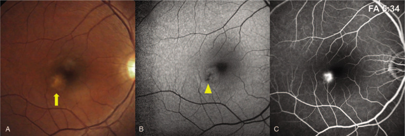
(A) Color fundus photography revealing a yellowish placoid lesion at the parafoveal area (arrow). (B) Fundus autofluorescence of the right eye revealing a circular area of hypo-autofluorescence compatible with retinal pigment epithelium destruction (arrow head). (C) Early fluorescein angiography revealing obvious dye leakage from the lesion at the parafovea.