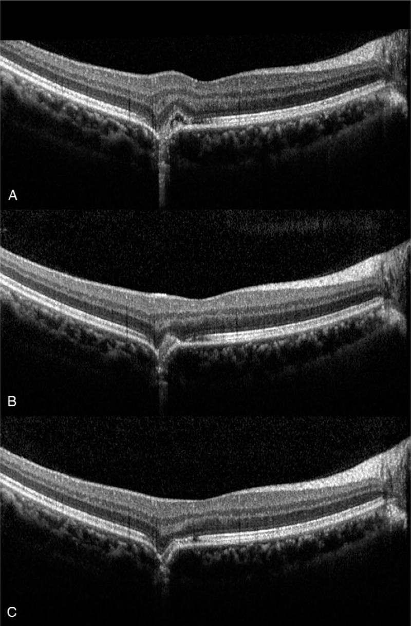
Figure 3.

(A) Optical coherence tomography (OCT) at the time of presentation revealing subretinal fluid and destruction of the retinal pigment epithelium and photoreceptor layer. (B) One week after intravitreal bevacizumab injection, the subfoveal fluid has decreased. (C) Four wk after intravitreal bevacizumab injection, OCT revealed marked improvement of the photoreceptor layer; however, excavated choroidal contour persisted. OCT = optical coherence tomography.
