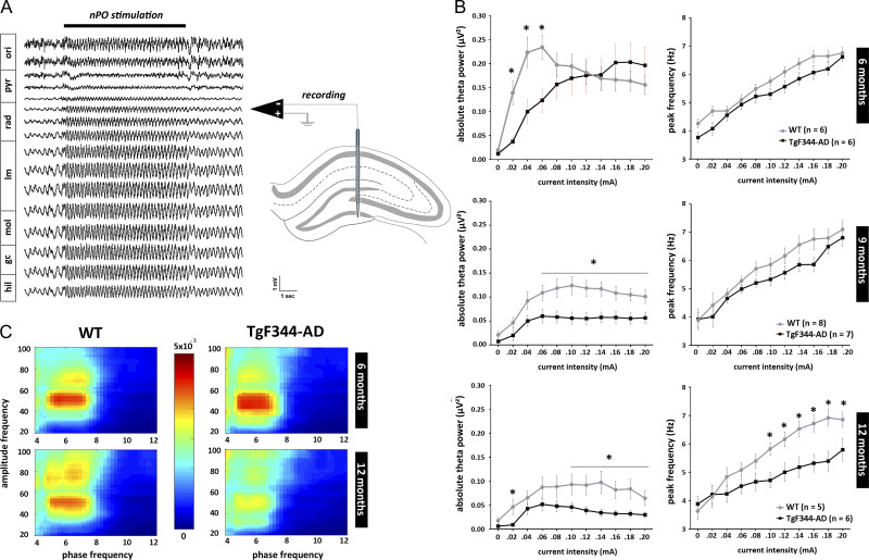
Figure 1.
Age-dependent disruption of elicited hippocampal oscillations in anesthetized TgF344-AD rats. High-frequency electrical stimulation of brainstem nucleus pontis oralis (nPO) induces highly regular theta oscillations in the hippocampal CA1 region (horizontal black bar marks the period of stimulation). Local field potentials were recorded using a multi-site silicon recording electrode implanted to span the dorsal hippocampus in urethane anesthetized rats. Absolute theta power (3–9 Hz) and peak theta frequency were determined for each stimulation period using fast Fourier transformation. The average of these measures calculated from the 3 channels showing strongest theta was used for each animal. (A) Typical stimulation-induced theta oscillation recorded from a WT rat. Approximate reconstruction of recording sites location is based on dorso-ventral position of the electrode and observations of highest theta power in lacunosum-moleculare (Buzsáki 2002). (B) Stimulus–response curves created by plotting the theta power and peak frequency over increasing stimulus intensities showed age-dependent decline in both theta power and frequency in TgF344-AD rats (*P < 0.05). (C) Elicited hippocampal theta and gamma oscillations showed high theta-phase gamma-amplitude coupling as expressed by Modulation index. Phase-amplitude coupling is significantly reduced in aged TgF344-AD rats (P < 0.05). Abbreviations: ori—str. oriens; pyr—pyramidal layer; rad—str. radiatum; lm—str. lacunosum-moleculare; mol—molecular layer; gc—granule cell layer; hil—hilus.

