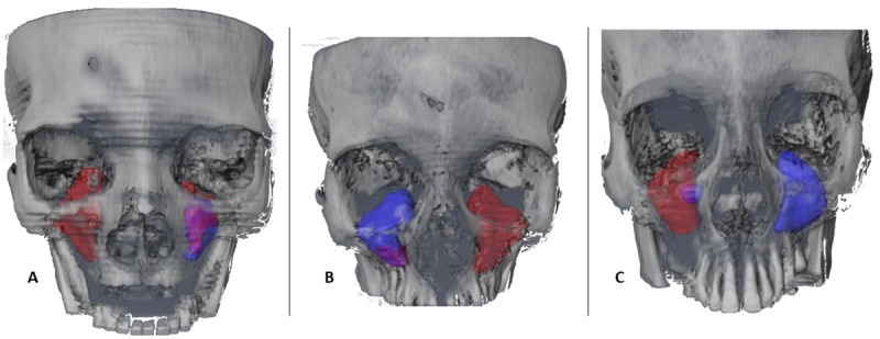
Fig 4. Volumetric images in frontal view.

Volumetric images in frontal view of the contour and the sinus occupation of 3 representative cases of the sample. In blue, the complete sinus volume, in red the volume which had been pathologically occupied, in purple the volume of residual superposition. A) CBCT16 Total right volume 119.6 mm3, occupied volume 119.6 mm3; total left sinus volume 71.5 mm3, occupied volume 24.6 mm3. B) CBCT10 total right volume 107.4 mm3, occupied volume 16.1 mm3; total left sinus volume 393.8 mm3, occupied volume 393.8 mm3. C) CBCT8 total right volume 241.7 mm3, occupied volume 209.6 mm3; total left volume 111.7 mm3, occupied volume 0 mm3.
