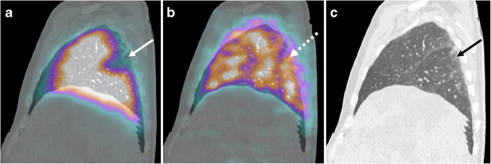
Fig. 1.
a Sagittal fused perfusion SPECT and CT (240 MBq of Pulmocis®). b Sagittal fused ventilation SPECT and CT (148 MBq of Technegas®). c Sagittal CT scan. Perfusion defect with pleural triangular base of the apical segment of the right lower lobe (white arrow) with normal ventilation in correspondence (white dotted arrow). CT scan shows sub-pleural ground glass predominant in the lower lobes (black arrow)

