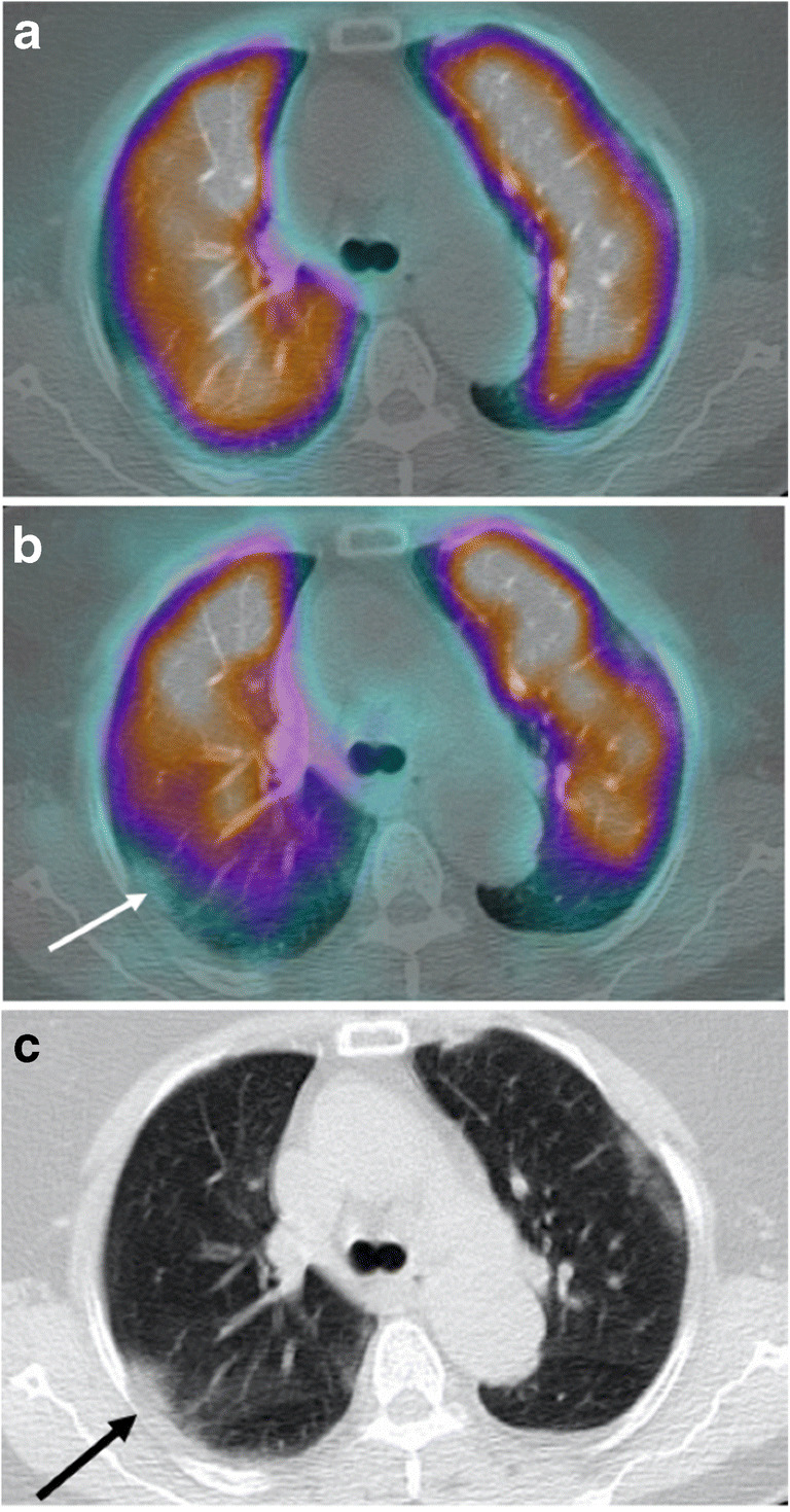
Fig. 5.

a Axial fused perfusion SPECT and CT (185 MBq of Pulmocis®). b Axial fused ventilation SPECT and CT (148 MBq of Krypton®). c Axial CT scan. Mismatched ventilatory defect in the posterior segment of the right upper lobe (white arrow), compared to ground glass on the scanner (black arrow)
