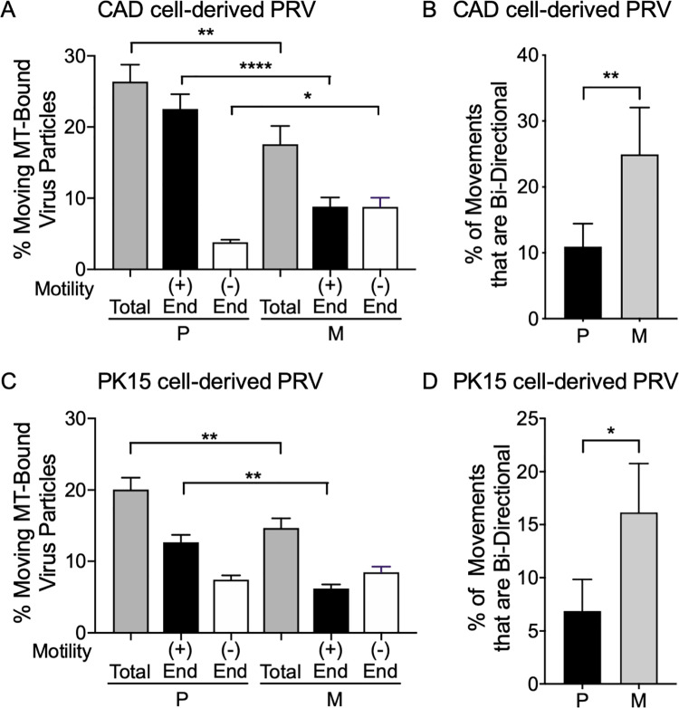
Fig 3. Loss of gE/gI-US9p reduces microtubule-mediated trafficking and increases frequency of minus end and bidirectional motion.
Float-up organellar fractions from P and M PRV-infected differentiated CAD or PK15 cells were bound to microtubules in a microscopic imaging chamber and their frequency and direction of in vitro motility determined. (A) Motile P and M PRV particles prepared from infected differentiated CAD cells. Total motile particles (grey bars) are expressed as a % of the number of viral particles bound to microtubules. Plotted values are the mean and standard deviation from the mean for 89 P and 82 M motile particles. In a separate experiment the motility of P and M PRV particles towards the plus end (+, black bars) or minus end (-, white bars) of directionally labeled microtubules was determined. Plotted values are the mean and standard deviation from the mean for 81 P and 54 M motile particles. Particles exhibiting bidirectional motion were excluded from this count. (B) P and M PRV particles derived from differentiated CAD cell float-up fractions that exhibited bidirectional motion on microtubules. Plotted values are mean and standard deviation from the mean for 9 P and 15 M motile particles. (C, D) Similar to (A, B) but testing motility of PRV particles in float-up fractions prepared from PK15 cells. Plotted values are mean and standard deviation from the mean for 99 P and 84 M motile particles (C) and 5 P and 13 M motile particles (D). * P≤ 0.05, ** P≤ 0.01, **** P≤ 0.0001.

