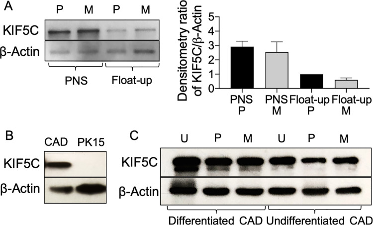
Fig 5. Western blots to demonstrate expression of KIF5C in infected and uninfected cells.
(A) Left of panel: Western blot of PNS and float-up fractions prepared from P or M PRV-infected differentiated CAD cells. Right of panel: Densitometric analysis of anti-KIF5C and anti-β−actin reactive bands from similar Western blots (data is the mean and range derived from two independently generated blots). (B) Western blot of PNS prepared from P PRV-infected PK15 or differentiated CAD cells. (C) Western blot of whole cell lysates prepared from differentiated or undifferentiated CAD cells that had been infected with P or M PRV strains or uninfected (U). In all panels the Western blot for β−actin provides a loading control.

