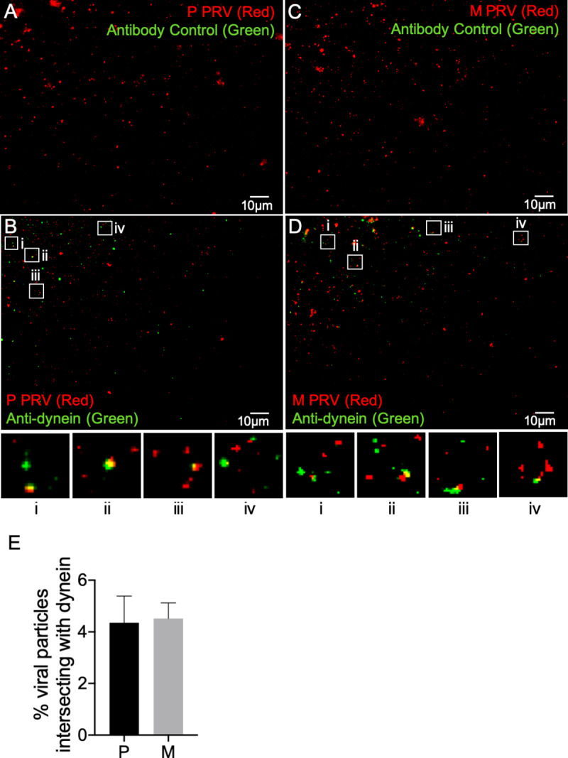
Fig 8. Loss of gE/gI-US9p does not affect the efficiency of dynein recruitment.

Float-up fractions from P or M PRV-infected differentiated CAD cells were immunostained with a mouse monoclonal anti-dynein antibody or the antibody omitted as control (indicated by text in each panel), imaged in the red (mCherry-tagged capsids) and green (anti-mouse antibody immunofluorescence) channels and images merged. (A, B) Fields of float-up particles from a P PRV infection stained in the absence or presence of anti-dynein IgG, respectively. (C, D) Fields of float-up particles from a M PRV infection stained in the absence or presence of anti-dynein IgG, respectively. Four boxed regions (i-iv) in panels B and D were magnified 3-fold and are shown at the bottom of each panel. The individual channel images used to construct the merged panels in A-D are shown in inverted black and white in S6 Fig and S7 Fig. (E) Numbers of P and M red fluorescent particles that exhibit anti-dynein staining as a percent of total particles in the field, derived from images similar to those in (B) and (D). Plotted values are mean and standard deviation from the mean for 7,914 P particles (imaged from 14 independent microscopic fields) and 7,932 M particles (imaged from 13 independent microscopic fields).
