FIGURE 3.

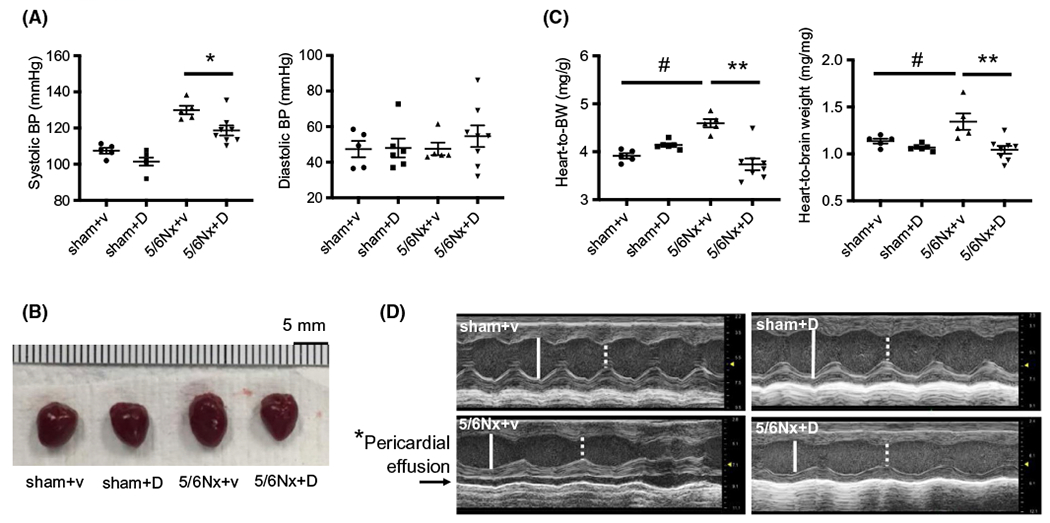
Inhibition of UT-A by dimethylthiourea (DMTU) ameliorated CKD-induced cardiac hypertrophy and improved heart function. A, Systolic and diastolic blood pressure at 8 weeks after completion of the CKD (5/6Nx) surgery. Blood pressure measurement of mice was performed by tail-cuff method. V = vehicle, D = DMTU treatment. B, Pictures of mouse hearts at sacrifice. C, Heart weight (mg) normalized by body weight (g) (left panel) and brain dry weight (mg) (right panel) at sacrifice. All data: mean ± SEM (N = 5-8), *P < .05, **P < .001, #P < .005 by one-way ANOVA. D, Echocardiography was performed on isoflurane anesthetized mice at 8 weeks of CKD. Images in each panel are M-mode images. White solid line—LVEDD: left ventricular endo-diastolic dimension; and white dash line—LVESD: left ventricular end-systolic dimension. Arrow in panel of 5/6Nx + vehicle denotes fluid space indicative of pericardial effusion. *P < .05 between CKD/+vehicle to the other three groups by Chi square analysis
