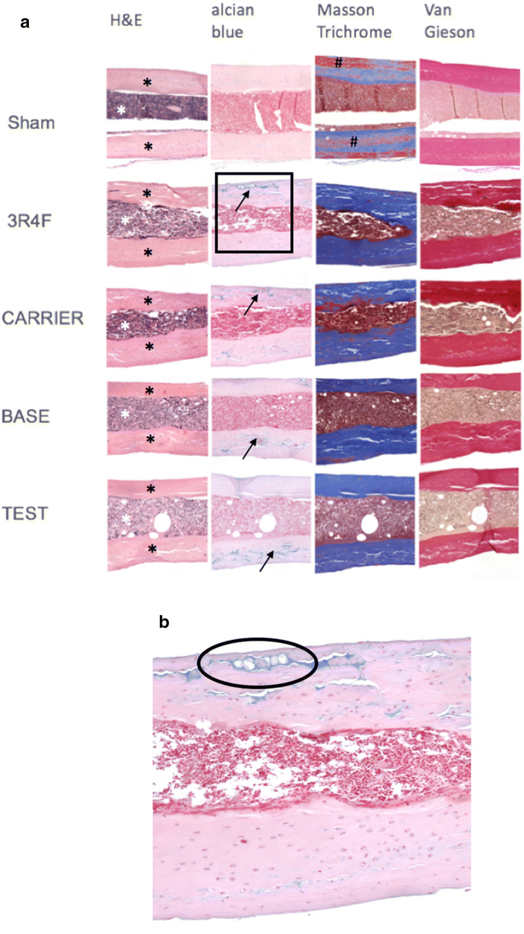
Fig. 5.
Bone morphology analysis by general histological analysis: a all groups were analyzed by histological staining. H&E staining provided a general overview of the cortical tissue (black stars) and cell-enriched intramedullary bone area (white star). Alcian blue staining depicted hypertrophic chondrocytes within cortical bone areas across all treated groups (black arrows). Masson trichrome staining showed red staining in cortical areas, mainly in the sham group (#). Cortical areas consisting of collagen were stained blue; red staining might show a variation in the types of collagen. Van Gieson staining revealed mineralized dark red areas in cortical bone, and mainly intramedullary cell cytoplasm was stained light brown. Additionally, in all treated groups, cortical bone areas revealed irregular contours facing the intramedullary area (white arrows). None of the mentioned cortical alterations (microcracks, irregular contours) were identified in Sham bones. b Enlarged section of a 3R4F-exposed sample stained with Alcian blue (black square in (a) depicting hypertrophic chondrocytes in a microcrack area (black circle)), providing evidence of endochondral bone repair. Sham: N = 4; 3R4F: N = 2; CARRIER: N = 3; BASE: N = 3; TEST: N = 3

