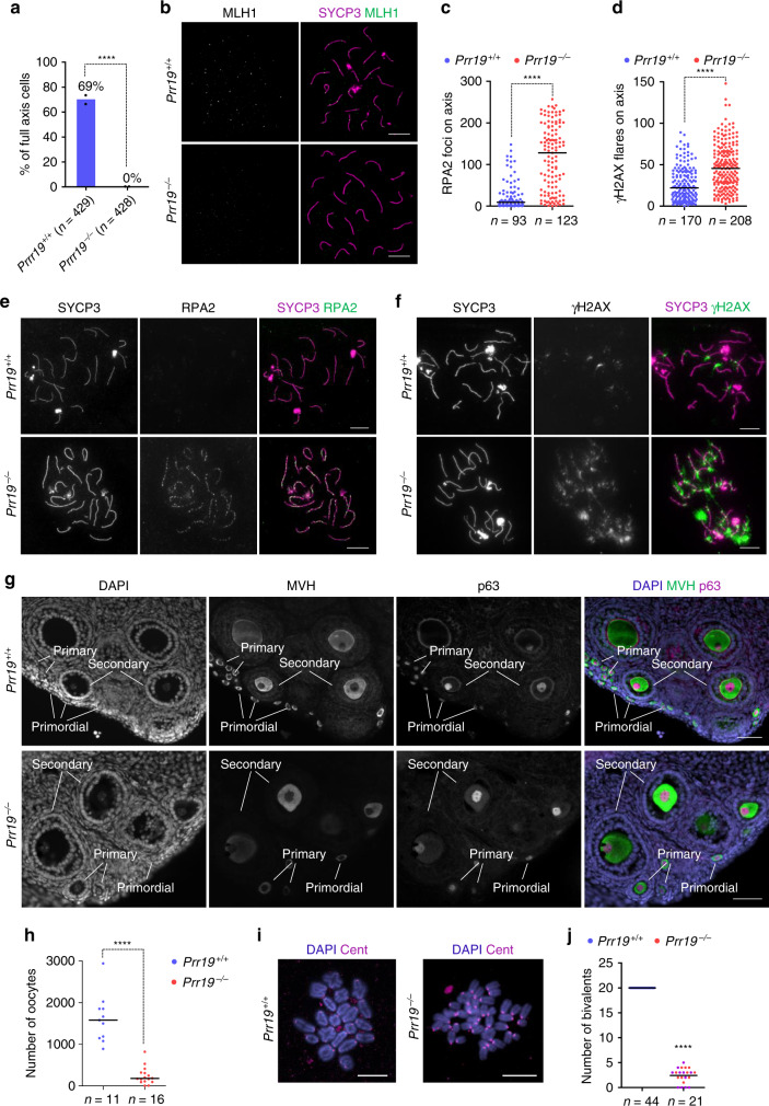
Fig. 6. Crossover formation and DSB repair are defective in Prr19−/− oocytes.
a Diagram shows percentages of oocytes with MLH1 foci in pools of oocytes that had fully formed axes in 18 dpc foetuses, datapoints and weighted averages of percentages are shown. n = numbers of analysed cells from two animals. Fisher’s exact test, P < 2.2E-16 (****). b, e, f Images show immunofluorescence of indicated proteins in nuclear spreads of (b) mid, (e) late pachytene or (f) early diplotene oocytes from (b) 18 dpc foetuses or (e, f) newborn mice. Bars, 10 µm. c, d Quantification of RPA focus numbers (c) and γH2AX flare numbers (d) in oocytes with fully formed axes from newborn mice. n = numbers of analysed cells from two mice; Mann–Whitney U test compared medians (bars) which are as follows in wild-type and Prr19−/−: RPA, 9 and 128 (P < 2.2E-16), γH2AX, 22 and 45.5 (P = 2.83E-14). g Oocytes were immunolabelled on ovary sections with MVH (cytoplasmic) and p63 (nuclear). DNA was labelled by DAPI. Primordial, primary and secondary follicles are indicated. Bars, 50 µm. h Quantification of oocyte numbers in ovary sections from 6- to 7-weeks-old females of indicated genotypes. Sums of oocyte numbers from every 6th sections of both ovaries of each mouse are shown. n = numbers of analysed animals; Mann–Whitney U test compared median numbers of oocytes (bars, wild-type, 1579 and Prr19−/−, 180.5), P = 1.53E-07 (****). i, j Oocytes were matured in vitro to metaphase I stage. i DNA was labelled with DAPI and centromeres (Cent) were detected by immunofluorescence on spreads of oocytes. Bars, 20 µm. j Quantification of bivalent numbers in spreads from matured oocytes. Numbers of analysed cells (n) and means (bars) are indicated. Note that mean bivalent number (2.4) is an underestimate in Prr19−/− oocytes, as we were able to identify full chromosome sets only in 10 out of 21 analysed Prr19−/− oocytes (red circles) due to a disorganised metaphase plate; more than half of the full chromosome set was detected in each of the remaining 11 oocytes (lilac circles). Statistics by one-tailed one-sample t test, P < 2.2E-16 (****). Source data are provided as a Source Data file.

