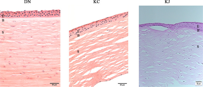
Figure 1.
Histology of patient and donor cornea sections. The figure shows similar retention of epithelial layers in DN and KC corneas, only KJ sample showed irregular and thickened epithelial cell layer in H&E staining of paraffin embedded sections. KC: keratoconus cornea from an African American patient; KJ: keratoconus cornea from a Middle Eastern patient; E: Stratified squamous epithelium; B: Bowman’s Layer; S: stroma; K: keratocytes.

