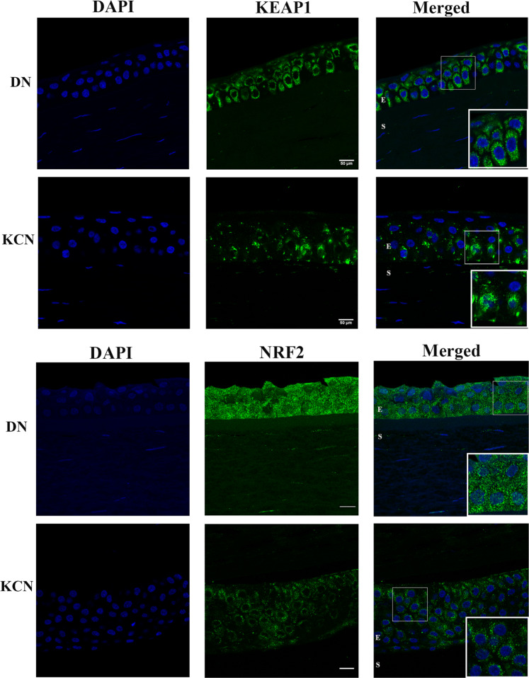
Figure 6.
KEAP1 and NRF2 immunostaining in DN and KCN corneas. (A) KEAP1 staining is decreased in KCN corneas, with focally increased staining in some basal epithelial cells (inset), whereas in DN corneas KEAP1 shows staining of all epithelial layers (inset). (B) NRF2 shows very little to no staining of KCN corneas and these were all cytoplasmic (inset), while DN sections show stronger staining of epithelial cells and some nuclear staining (inset) DAPI nuclear staining shown in blue. IF staining of additional KCN and DN cornea sections are shown in Supplemental Fig. S4. Scale bar: 50 µm.

