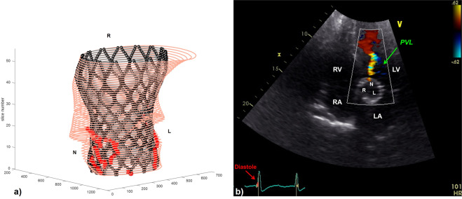
Figure 5.
(a) Matlab elaboration of the FE results for patient C-8, highlighting the presence of PVL in the non-coronary cusps (NCC) of the valve (red asterisks). (b) Transthoracic echocardiographic 5 chamber view for the same patient: one jet of paravalvular regurgitation (PVL) is showed by the green arrow in the non-coronary cusp. The ECG shows that the image was taken during diastole, i.e during the bioprosthetic valve closure. RV = right ventricle, RA = right atrium; LA = left atrium; LV = left ventricle; R = right coronary cusp; L = left coronary cusp; N = non-coronary cusp.

