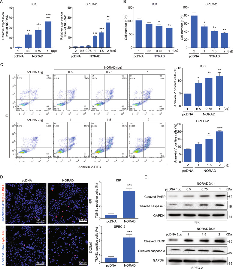
Fig. 2. NORAD promotes apoptosis in EC cells.
a qRT-PCR analysis for the expression level of NORAD in ISK and SPEC-2 EC cell lines with different doses transfection of NORAD, in comparison with the empty vectors. b Cell-counting assays for the control and ectopic NORAD-expressing ISK and SPEC-2 cells after 48 h transfection. c Increased percentage of apoptosis in the ectopic NORAD-expressing EC cells after 48 h transfection via FACS analysis. d TUNEL assays for apoptotic cells in the control and NORAD-expressing EC groups (left). Statistics of the TUNEL-Cy3 positive cells are shown (right). Scale bar, 100 μm. e Activated expression of cleaved PARP and cleaved caspase-3 was visualized by western blot. The results were determined from triplicates, and the error bars represented as the mean ± SD, *P < 0.05, **P < 0.01, ***P < 0.001. TUNEL TdT-mediated dUTP Nick-end labeling, GAPDH glyceraldehyde-3-phosphate dehydrogenase.

