
FIGURE 2.
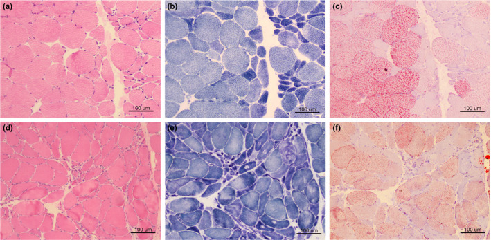
The myopathological features of the two patients. Muscle biopsy shows grouping of small angular atrophic fibers on HE stain, suggesting a neurogenic process (a: case 1; d: case 2). NADH stain reveals dark angular fibers and target fibers (b: case 1; e: case 2). Numerous lipid droplets aggregate in the relatively hypertrophy fibers on ORO stain (c: case 1; f: case 2)