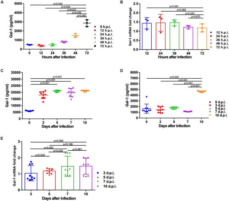
FIGURE 2.
Expression of Gal-1 protein and mRNA after infection with H1N1pdm09 virus. (A) Gal-1 protein level in the supernatant of A549 cells at 0, 12, 24, 36, 48, and 72 h after infection with H1N1pdm09. (B). The fold change of Gal-1 mRNA level in A549 cells at 12, 24, 36, 48, and 72 h after infection with H1N1pdm09 (n = 3). (C,D) Gal-1 protein level in mouse BALF (C) and serum (D) at 0, 3, 5, 7, and 10 days after infection with H1N1pdm09. (E) The fold change of Gal-1 mRNA level in mouse lung homogenates at 3, 5, 7, and 10 days after infection with H1N1pdm09 (n = 9). Representative data shown from three independent experiments. P-values are indicated in the panels. BALF: bronchoalveolar lavage fluid.

