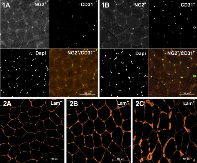
FIGURE 1.

Representative skeletal muscle cross sections displaying immunoreactivity for NG2 (red), CD31 (green), and 4’,6-diamidino-2-phenylindole (DAPI) (nuclear stain) (1A,1B) and Laminin (2A–2C). 1A,1B: Samples are from blood flow restriction exercise (BFRE) at baseline (Pre, 1A) and 8 days into the intervention (Mid8, 1B). Note the increase in the area of CD31C structures and myofibers as well as a similar number of CD31C structures, suggesting a stable capillary density (CD) and an increase in capillary/fiber and capillary cross-sectional area. 2A: Normal laminin morphology, baseline (Pre); 2B/2C: lowly/highly increased perivascular laminin immunoreactivity relative to baseline (2A).
