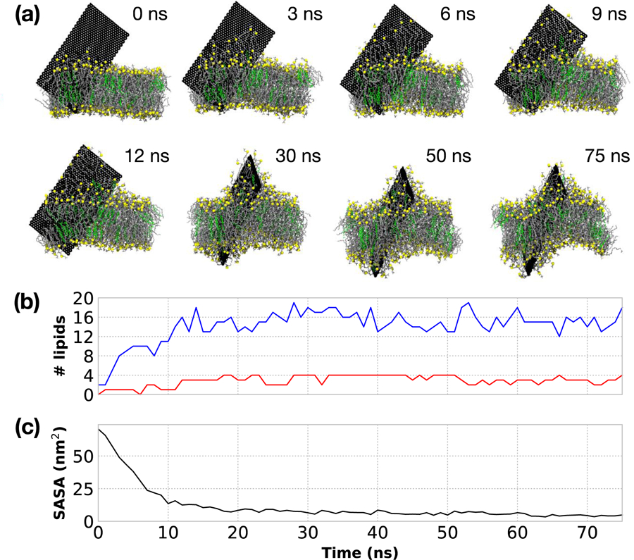
Figure 3:

(a) Simulation snapshots of the extraction of lipids for a 5 nm ×10 nm GNF. The GNF surface is fully coated by a single layer of lipids ~15 ns. After which additional lipids are extracted to further coat the lipids already on the surface of the GNF, providing additional shielding of the lipid tails, resulting in significant membrane disruption by the end of the simulation time considered (75 ns). Phosphorus shown in yellow, DOPC tails in grey, CHOL colored green. Water is not shown for clarity. The camera location is changed (i.e., rotated) for the final three snapshots to highlight the further extraction and disruption of the bilayer; the position of GNF itself does not change during the course of the simulation due to the applied restraints. (b) Quantification of the number of lipids extracted from the bilayer. DOPC shown in blue and CHOL shown in red. (c) Calculation of the surface accessible solvent area of the GNF as a function of time.
