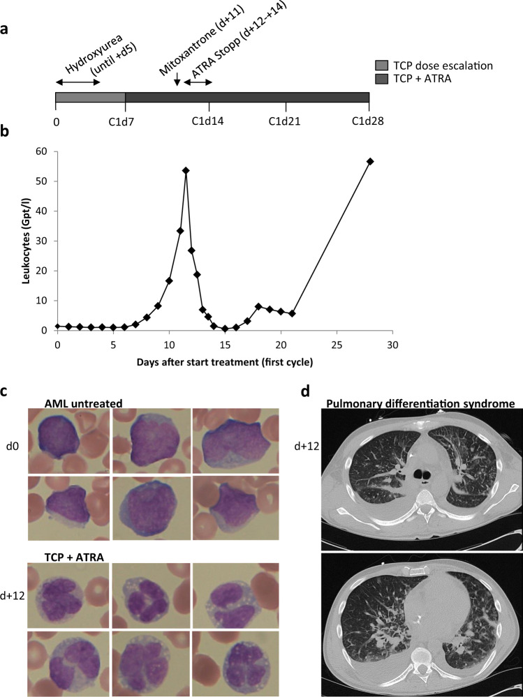
Fig. 4. Myeloid differentiation syndrome in a patient in the TCP/ATRA trial.
a Clinical course of the patient (05). b Numbers of leukocytes in the peripheral blood during the first cycle. c Cell morphology of AML cells in the peripheral blood on day one and day +12 of the first cycle. Peripheral blood smears are stained with Pappenheim and shown at a magnification of 63×. Treatment with TCP and ATRA leads to morphological signs of differentiation of blasts into polynuclear leukocytes. d Axial CT-scans on day +12 show centrilobular nodules and ground-glass opacity in both lungs with predominantly small spotted alveolar infiltrates and discrete interstitial thickenings. Further pleural effusions on the right more than on the left.

