Fig. 6.
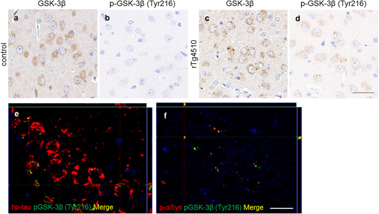
Immunohistochemistry for GSK-3β in the amygdala of rTg4510 and control mice. The cytoplasmic uniform distribution of GSK-3β was found in both mice (a, c). GSK-3β-positive grains were found in rTg4510 mice (c). P-GSK-3β (Tyr216)-positive cells were found in rTg4510 mice (d), but not in control mice (b). Double-labeling immunofluorescence in the hippocampus of rTg4510 mice revealed that p-GSK-3β (Tyr216)-positive grains were localized in hp-tau-positive cells (e). The majority of p-GSK-3β (Tyr216) grains did not co-localize with hp-tau aggregates (e). P-αSyn-positive aggregates localized in p-GSK-3β (Tyr216)-positive cells (f). p-αSyn-positive aggregates co-localized with p-GSK-3β (Tyr216)-positive grains (f). The reactions to hp-tau (e) and p-αSyn (f) are shown in red, and those to p-GSK-3β (Tyr216) (e, f) in green. Scale bars: 20 μm (a-d); 50 μm (e, f)

