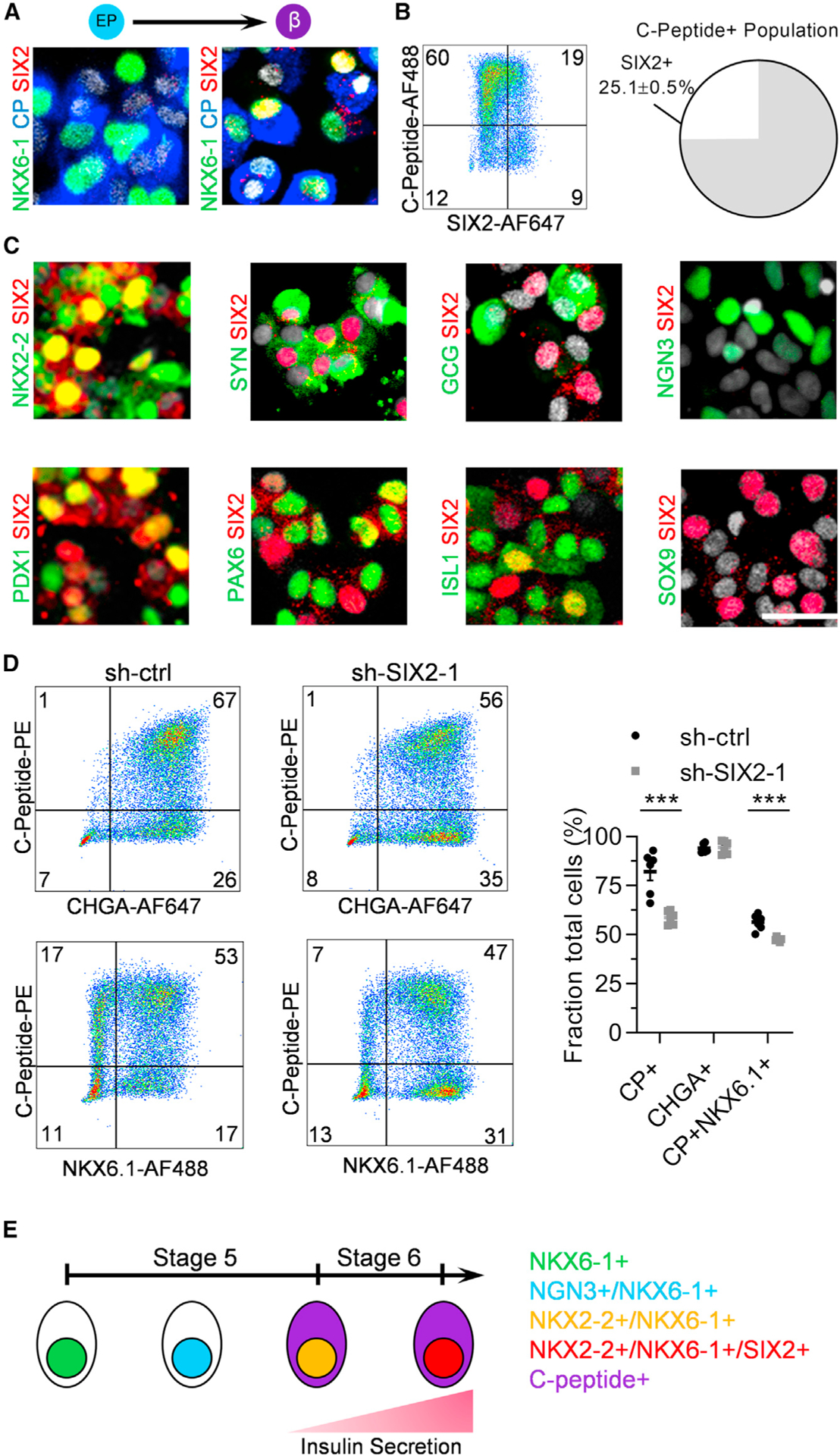
Figure 2. Subtypes of Differentiated Stage 6 Cells Express SIX2.

(A) Immunostaining of SIX2 with the β cell markers NKX6–1 and C-peptide at the end of stages 5 (left) and 6 (right).
(B) Flow cytometric quantification of co-expression of C-peptide with SIX2. n = 4.
(C) Immunostaining of SIX2 with a panel of pancreatic markers at the end of stage 6 with the exception of NGN3/SIX2, which was stained 3 days into stage 5.
(D) Flow cytometric quantification of stage 6 cells staining for C-peptide, NKX6.1, and chromogranin A using sh-ctrl and sh-SIX2–1 transduced cells. n =6. ***p < 0.001 by 2-way unpaired t test.
(E) Schematic summary of marker progression in stages 5 and 6. Scale bar, 25 mm. Error bars represent s.e.m. See also Figure S2.
