Fig. 1.
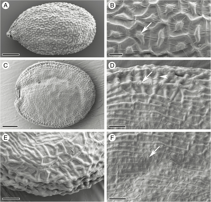
Cryo-scanning electron micrographs of mature seed surface structure. (A, B) A. thaliana seed (A) and surface topography (B) consisting of columella (arrow) and anticlinal cell walls of epidermal oi2 cells. (C–F) C. hirsuta seed (C) and surface topography (D–F) characterized predominantly by anticlinal cell walls of the sub-epidermal seed coat layer oi1 (arrowhead, D), especially at the seed margin (E). The columella and anticlinal cell walls of oi2 cells are still visible (arrow, D, F), especially on the flat seed surfaces (F). Scale bars = 100 µm (A), 20 µm (B), 200 µm (C) and 50 µm (D–F).

