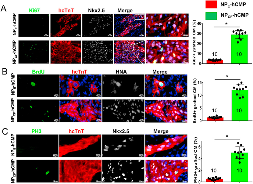
Fig. 4.
hiPSC-CM cell cycle activity and proliferation were greater in NPCF-hCMPs than in NPE-hCMPs four weeks after transplantation into infarcted mouse hearts. (A-C) Engrafted hiPSC-CMs in the NPCF-hCMPs and in the NPE-hCMPs were identified in mouse hearts at week 4 after MI induction and treatment via immunofluorescent staining for the expression of hcTnT and HNA or hNkx2.5; then, the proportion of hcTnT-hNkx2.5 double-positive cells (A and C) or hcTnT-HNA double-positive cells (B) that also stained positively for (A) Ki67 (a proliferation marker) (B) BrdU incorporation (an S-phase marker), and (C) PH3 (an M-phase marker) was determined and expressed as a percentage. The number of animals evaluated for each treatment group is displayed at the base of the corresponding column. Five randomly selected viewing fields were evaluated per section and at least six sections were evaluated per animal. *p < .01, two-tailed Student’s t-test.

