
Fig. 3. Hypoxia signaling inhibitors prevent DUX4-induced cell death.
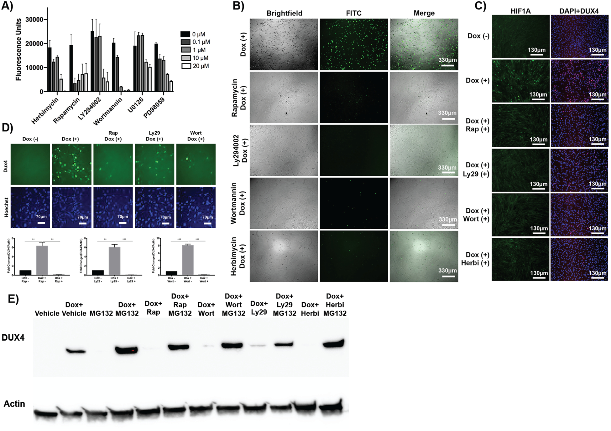
(A) Dose-response curves for known chemical inhibitors of hypoxia signaling. Cell death was quantified using a caspase 3/7 luminescence assay. (B) Fluorescence imaging analysis of DUX4-induced apoptosis in MB135-DUX4i cells incubated with PI3K/Akt/mTOR inhibitor compounds. Green cells are caspase-3/7 positive. (C) Fluorescence imaging analysis of hypoxia in MB135-DUX4i cells cultured with inhibitor compounds; HIF1A (green), DUX4 (red), DAPI (blue). (D) Immunofluorescence (P2B1 antibody, top) and quantification of western blot (E55 antibody, bottom) of DUX4 protein expression in the presence of signaling inhibitors in MB135-DUX4i cells. (E) Western blot for DUX4 protein with MG132 proteasome inhibition in the presence of inhibitors in MB135-DUX4i cells.