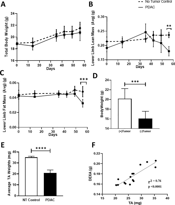
Fig 3. Longitudinal quantification of lean mass via DEXA demonstrates commencement of PDAC-related muscle wasting within 38 days of tumor implantation, which cannot be detected by assessment of total body weight.
Mice were randomized to two groups prior to orthotopic tumor injections: 1) NTC; (n = 5) and 2) PDAC tumor bearing; (n = 5). Longitudinal DEXA and total body weight (TBW) measurements were performed as described in Materials and Methods. (A) Longitudinal TBW is presented for the NTC and PDAC mice. Longitudinal lower limb (B) lean and (C) fat mass was determined by DEXA. Data is presented as mean ± SD by two-way ANOVA for repeated measures (the interaction between PDAC vs. NTC and time was significant at day 56 for lean mass and fat mass **p = 0.002 and ***p = 0.0003, respectively), Tukey’s multiple comparison test. (D) The total body weight (TBW) (g) and (E) TA weights (mg) of the PDAC bearing mice was determined on the day of sacrifice, and the data are presented as TBW (+ tumor) before primary tumor harvest and net weight (- tumor) after primary tumor harvest for each mouse with the mean ± SD, Student t-test revealed a significant difference (***p = 0.0005) (****p<0.0001) between both bodyweight and TA weight, respectively. (F) A Pearson correlation analysis was performed on a separate and larger cohort of PDAC tumor bearing mice (n = 15) to determine the relationship between DEXA lean mass and TA weight. The analysis confirmed a significant (p < 0.0001) relationship between the two measures with r2 = 0.76. Data are expressed as mean ± SD.

