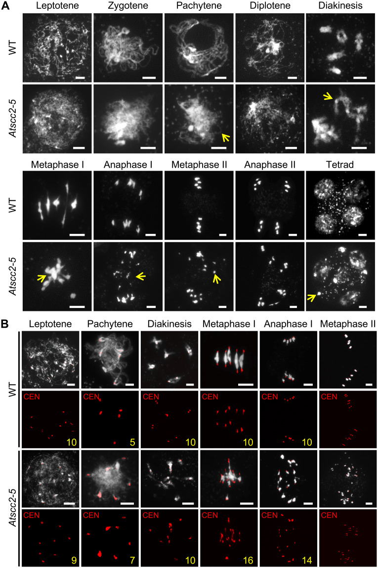
Fig 2. Chromosome morphology of Atscc2-5 and WT male meiocytes.
(A) Chromosome spreads of WT and Atscc2-5 male meiocytes stained by DAPI. Yellow arrows indicate the asynaptic chromosomes, univalent, abnormal chromosomal entanglements or fragments in Atscc2-5. Bar = 5 μm. (B) Fluorescence in situ hybridization (FISH) of WT and Atscc2-5 chromosomes using a centromere probe. Yellow numbers indicate the number of centromeres in the meiocytes. Bar = 5 μm.

