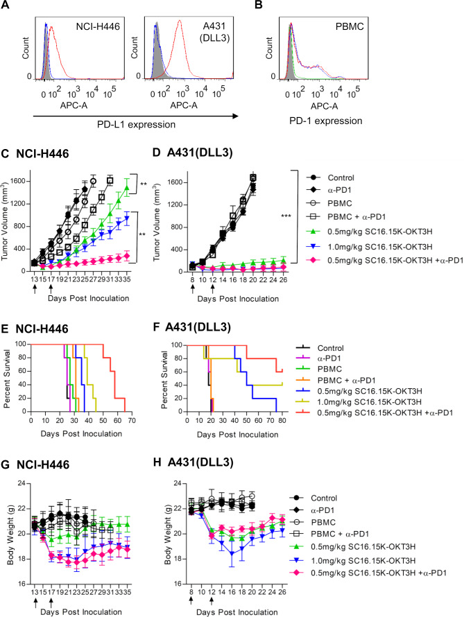
Figure 4.
In vivo efficacy testing of the delta-like 3 (DLL3) bispecific antibody. (A) PD-L1 expression on H446 and A431 (DLL3) cells. One million of cells was stained with PD-L1 rabbit monoclonal antibody, followed by detection with APC-conjugated goat-anti-rabbit IgG. Shaded area, secondary antibody staining; blue curve, isotype control (pooled rabbit IgG) staining; red curve, PD-L1 antibody staining. (B) PD-1 expression on the DLL3 bispecific antibody-stimulated peripheral blood mononuclear cells (PBMC) cells. H446 (red curve) and A431(DLL3) cells (blue curve) were incubated with PBMC for 48 hours in the presence of 100 ng/mL DLL3 bispecific antibody, followed by flow cytometry to analyze PD-1 expression. Shaded area, unstimulated PBMC cell staining; green curve, isotype control (pooled human IgG) staining of H446-stimulated PBMC. (C) Tumor growth curve of native small cell lung cancer cell line NCI-H446. Five million cells were subcutaneously inoculated in each NSG mouse. After the tumor formed and reached a size of 100–200 mm3, treatment was started. Bispecific antibody alone (0.5 mg/kg or 1.0 mg/kg body weight), or in combination of anti-PD-1 antibody (in-house made scFv-hFc format, 5.0 mg/kg) were tested. Ten million unstimulated human PBMC were intraperitoneally given immediately before the first intravenous delivery of the antibody. Arrows indicated the injection time point of the bispecific antibody. The PD-1 antibody was intravenously given once every week since the start of the treatment. Both tumor volume and body weight were measured every two or 3 days. **p<0.01, calculated using a Student’s paired t-test (two-tailed), ***p<0.001. (D) Tumor growth curve of A431 (DLL3), an artificial A431 cell line that was forcefully overexpressing DLL3. ***p<0.001, calculated using a Student’s paired t-test (two tailed). (E) The survival curves of the H446 mouse model treated with the bispecific antibody. (F) The survival curves of the A431 (DLL3) mouse model. (G) Body weight of the H446 mouse model treated with the bispecific antibody. (H) Body weight of the A431 (DLL3) mouse model.

