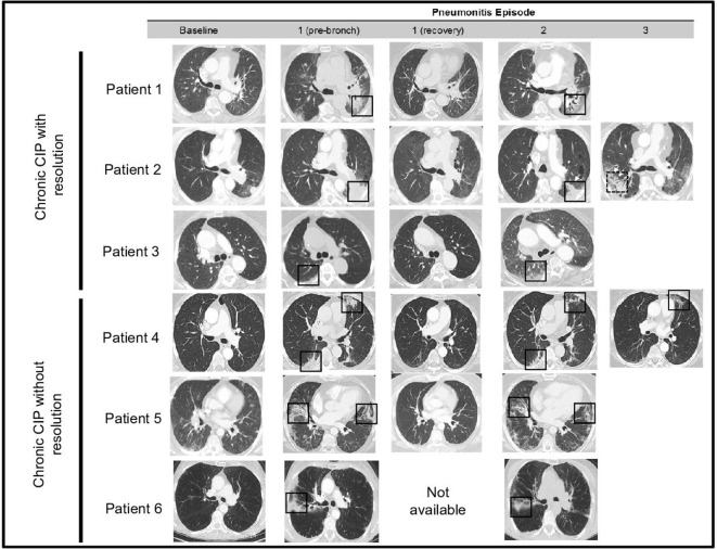
Figure 1.
Serial radiological imaging in patients with chronic immune checkpoint inhibitor pneumonitis. Representative CT images at baseline—1: (pre-bronch): prior to first bronchoscopy, 1 (recovery): after treatment of first pneumonitis episode, 2: second episode of pneumonitis, 3: third episode of pneumonitis. Solid boxes show initial sites of pneumonitis and recrudescence, with radiographic pattern in keeping with acute lung injury or organizing pneumonia. Dotted line box highlights a new area of airspace opacity.

