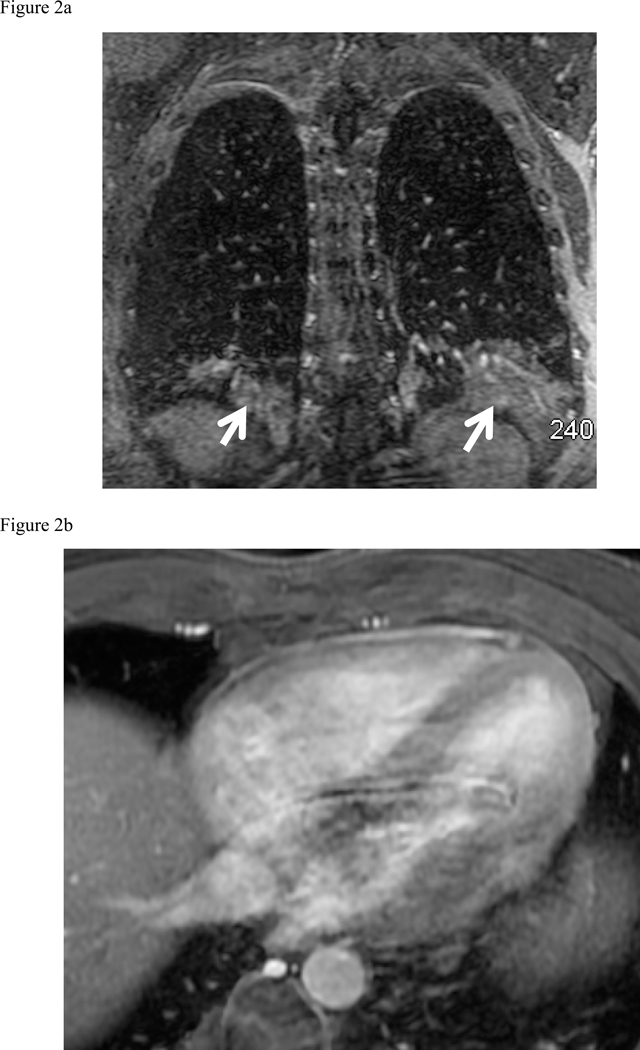
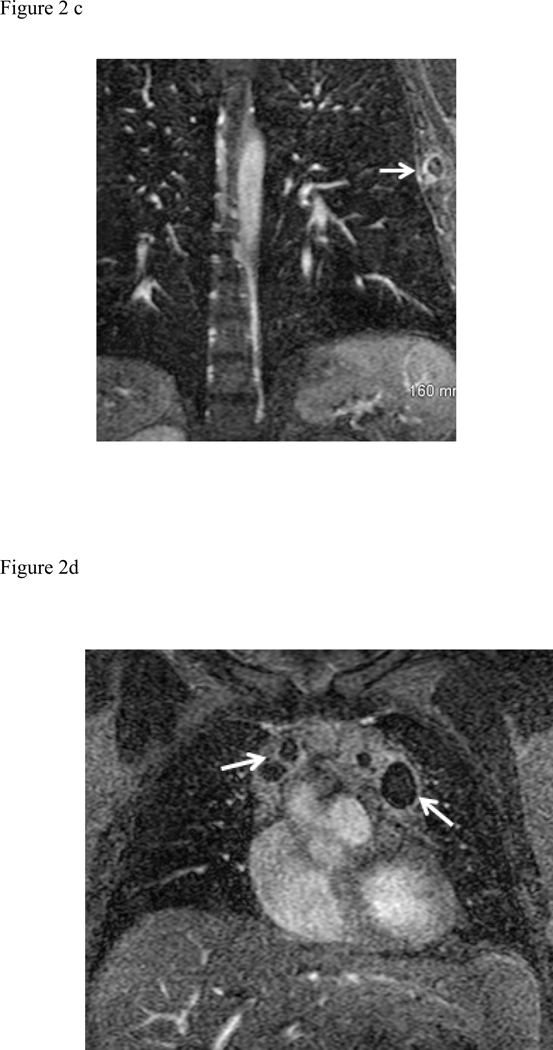
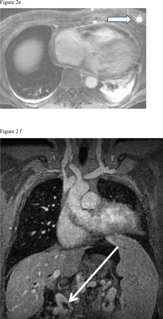
Fig. 2:
Composite collection of Type 1 actionable findings. (a) Pneumonia- shown on the MRA delayed phase in coronal plane shows consolidation in both lower lobes (white arrows), (b) Pericarditis- enhancing visceral and parietal pericardial tissue (arrow) shown on the MRA delayed phase of contrast enhancement (c) Left lateral 6th rib fracture (arrow) shown on the MRA delayed phase (d) MRA delayed phase in coronal plane shows multiple enlarged necrotic lymph nodes - Hodgkin’s disease (white arrows), (e) Left breast mass at the post contrast T1-weighted images (arrow), (f) Portal vein thrombosis (arrow) shown on the CE-MRA delayed phase of contrast enhancement.


