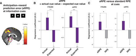
Fig. 3. Neural representation of our computational model’s aRPE signals.

(A) The ventral tegmental area and substantia nigra (VTA/SN) and medial posterior parietal cortex (mPPC) BOLD positively correlated with the model’s aRPE at the time of advanced information cue presentations [VTA/SN, P < 0.05 FWE small volume correction (39); mPPC, P < 0.05 whole-brain FWE, cluster-corrected at P < 0.001]. Voxels at P < 0.005 (uncorrected) are highlighted for display purposes. (B) Our confirmatory analysis shows that both the VTA/SN and the mPPC show paradigmatic correlations with aRPE. At the time of advanced information cue presentations, BOLD in the VTA/SN and the mPPC positively correlated with the model’s actual cue value signal and negatively with the model’s expected cue value signal, indicating that both regions express canonical prediction errors. The difference was significant in the VTA/SN (P < 0.001, permutation test) and in the mPPC (P < 0.001, permutation test). The positive correlation with cue outcome values and the negative correlation with expected values were all significant (received cue value, P < 0.01 for the VTA/SN and the mPPC by t test, t38 = 3.24 and t38 = 3.40; expected cue value, P < 0.01 for the VTA/SN and P < 0.001 for the mPPC by t test, t38 = 2.82 and t38 = 4.37). (C) Our confirmatory analysis shows that both regions express stronger correlations with our model’s full aRPE than with standard prediction error with discounted reward (RPE) at advanced information cues. The difference was significant between the VTA/SN and in the mPPC cluster (P < 0.05, permutation test). ***P < 0.001, **P < 0.01, and *P < 0.05.
