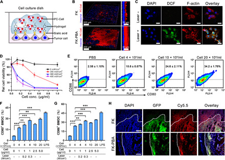
Fig. 3. PC-Cell@gel induced PDT and DC maturation in vitro.

(A) Scheme illustrating the gelation of FK-PBA in vitro. Seeded B16-OVA cells were stained by Hoechst, fixed, and sequentially treated with FK-PBA and PC-Cell in 1 mM Na2CO3 solution. (B) 3D construction of PC-Cell@gel on the surface of B16-OVA cells (right). Scale bars, 200 μm. Side view of the 3D structures (left). Scale bars, 50 μm. (C) PC-Cell@gel induced ROS production in B16-OVA cells. Scale bars, 20 μm. (D) Laser power density–dependent phototoxicity of PC-Cell@gel. (E) Cell@gel promoted BMDC maturation following increased vaccine cell concentrations. (F) Percentage of CD11c+CD80+ BMDCs and (G) CD11c+CD86+ BMDCs after incubated with PC-Cell@gel or laser-pretreated PC-Cell@gel. LPS, lipopolysaccharide. (H) Mice with partially resected CT26-GFP tumors were first locally treated with FK-PBA-Cy5.5 or FK-Cy5.5 at 5 mg/ml, respectively. Four hours later, 1 mM Na2CO3 solution was sequentially injected. Tissues in surgical bed were collected and sliced for CLSM imaging at 24 hours. Scale bars, 200 μm. Data are means ± SD (n = 3). **P < 0.01, ***P < 0.001.
