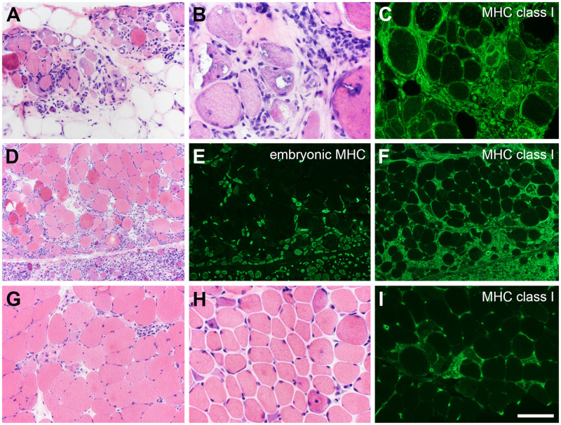
FIGURE 10.
Myositis and immune-mediated necrotizing myopathy (IMNM). The more common forms of inflammatory myopathy are readily distinguished by hematoxylin and eosin staining and immunofluorescence. (A–C) In this patient with inclusion body myositis, the biopsy manifests lymphocytic infiltrates, endomysial fibrosis, fatty replacement, rimmed-vacuoles, and diffuse expression of myosin heavy chain (MHC) class I. (D–F) A perifascicular pattern of myonecrosis, regeneration, and MHC class I expression define this example of dermatomyositis spectrum disorder. Embryonic myosin heavy chain is a marker of regeneration (E). IMNM is not easily distinguishable from other types of necrotizing myopathy (G–I). The patient illustrated in panels G and I became symptomatic while taking a statin. The patient shown in panel H is statin-naïve and was followed for several years with a clinical diagnosis of limb-girdle muscular dystrophy. Both patients have highly elevated anti-HMGCR antibodies. The pattern of MHC class I expression (I) closely follows the pattern of myonecrosis. The scale bar in panel I is 50 µm for panels B and H; 100 µm for panels A, C, G, and I; 200 µm for panels D–F.

