FIGURE 7.
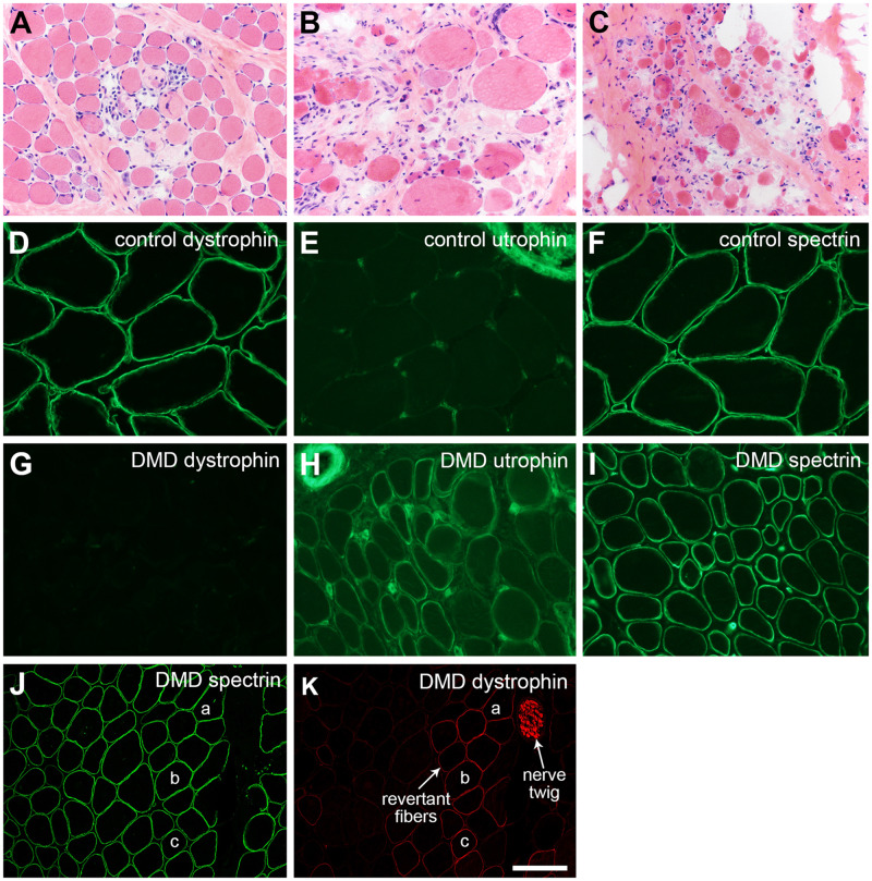
Duchenne muscular dystrophy (DMD). The severity of dystrophic pathology may vary extensively depending on the DMD mutation, the age of the patient at the time of biopsy, and the muscle biopsied. (A) Early, mildly dystrophic pathology with active myonecrosis and regeneration is illustrated in panel. (B, C) Later (chronic), more severe dystrophic pathology is shown. Immunofluorescence evaluation of DMD muscle shows a loss of dystrophin (cf. panels D and G) and upregulation of utrophin (cf. panels E and H). Utrophin positivity in the upper right corner (E) is a nerve twig and in the upper left corner (H) is a small artery. Endomysial capillary endothelia are normally positive as seen in both E and H. Spectrin immunostaining assures that the DMD muscle fibers are non-necrotic and free of nonspecific proteolytic degradation (F, I). Dystrophin-positive revertant fibers and Schwann cells are commonly encountered in muscle biopsies from DMD patients (K). The spectrin immunofluorescence of the same cryosection (staining performed by dual label methodology) is shown in panel J; muscle fibers “a,” “b,” and “c” are the same in each panel (J, K). The scale bar in panel K is 50 µm for panels D–H and I; 100 µm for panels A–C, J, and K.

